
Lack of CLN3 harms the process of survival for cells under stress: Study
Missing in juvenile Batten, CLN3 protein is 'important' for cellular stress response
Written by |


A lack of the CLN3 protein — the underlying cause of juvenile Batten disease — impairs the function of stress granules (SGs), which are formed to promote cell survival under stressful conditions, according to a cell-based study.
CLN3-deficient cells also had lower metabolic rates and were impaired in their ability to handle additional stressors. Such defects increase cellular stress, making cells with high energy demands, such as nerve cells, more vulnerable to damage.
“With a growing interest in SG-modulating drugs for the treatment of neurodegenerative diseases,” researchers wrote, “novel insights into the molecular basis of [juvenile] Batten disease may reveal avenues for disease-modifying treatments for this debilitating childhood disease.”
The study, “The Batten disease protein CLN3 is important for stress granules dynamics and translational activity,” was published in the Journal of Biological Chemistry.

Mutations in CLN3 gene lead to juvenile Batten disease
Batten disease is a group of neurological conditions caused by inherited mutations that impair lysosomes, which are the cell’s recycling centers. These defects lead to the build-up of waste products to toxic levels, resulting in tissue damage, especially to nerve cells.
Mutations in the CLN3 gene, which encodes a protein found in the membrane of lysosomes, lead to juvenile Batten, also known as CLN3 disease.
Previous research to understand CLN3 protein’s function, and its role in disease, suggests it regulates several degradation and recycling processes, including those of fat molecules used to make cell membranes.
CLN3’s loss has also been linked to impaired energy metabolism. This includes the altered function of energy-producing mitochondria and higher oxidative stress, a type of cellular damage caused by the build-up of damaging reactive oxygen species generated by metabolism.
Because nerve cells have high energy demands, they are especially vulnerable to metabolic stresses.
In response to such stress, cells usually shut down normal protein production and reorganize stalled proteins and RNA molecules into SGs. Rapidly assembled around a scaffold protein called G3BP1, SGs modulate several signaling pathways to promote survival until the stress clears.
Evidence shows persistent SGs that do not disassemble after stress are associated with several neurological conditions. These data prompted a team of researchers in the U.K. to investigate the relationship between CLN3 and the SG pathway using lab-grown human cells genetically modified to lack the CLN3 gene (CLN3-KO).
Initial experiments confirmed impaired energy metabolism in CLN3-deficient cells relative to healthy, unmodified cells, which were used as controls.
The drop in metabolic rate with the addition of a stressor molecule called sodium arsenite (NaArs) was “more profound” in CLN3-KO cells, the researchers wrote, showing these cells have “an impaired ability to handle additional stressors and start with a lower metabolic turnover in the absence of stressors.”
This was “indicative of impaired mitochondrial function,” they added.
Protein production within CLN3-deficient cells was also impaired, as were signaling networks involved in sensing and responding to stressors.
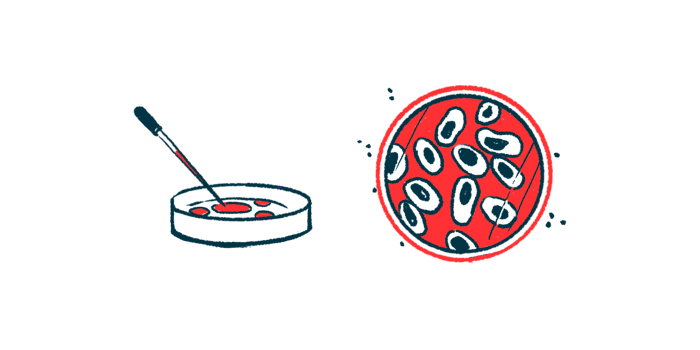
Cells were then exposed to NaArs to trigger the assembly of SGs, which was detected by G3BP1 protein staining. Small and medium SGs formed within five minutes.
However, after 20 minutes, there were 50% fewer large SGs in CLN3-KO cells than in control cells, “suggesting an impairment in the maturation of SGs in the later stages of SG assembly,” the team wrote.

‘CLN3 may be important for recovery from and adaptation to oxidative stress’
Unlike control cells, a significant number of small and medium SGs persisted in cells lacking CLN3 three hours after removing the NaArs stressor. This suggested that “the clearance of NaArs-induced SGs is less efficient in [CLN3-KO], and that CLN3 may be important for recovery from and adaptation to oxidative stress,” the researchers wrote.
By comparison, genetically modifying cells to lack the CLN5 protein — an underlying cause of late infantile Batten disease — did not affect SG assembly or disassembly.
To validate these findings, human connective tissue cells, called fibroblasts, were collected from juvenile Batten patients and unrelated healthy donors. These fibroblasts were grown in the lab and exposed to NaArs.
While most SGs in control cells disassembled after 90 minutes, patient fibroblasts had persistent SGs up to two hours post-recovery, “supporting a more general mechanism linking CLN3 and delayed SG recovery,” the researchers wrote.
Finally, the activity of the gene that encodes the G3BP1 scaffold protein was significantly lower in CLN3-KO cells and those derived from patients than in their respective controls. This difference resulted also in lower levels of the G3BP1 protein.
“Our study paves the way for further investigation in patient-derived cells to explore the link between lysosomes, SG dynamics and modulation in CLN3 disease,” the researchers concluded.

Leave a comment
Fill in the required fields to post. Your email address will not be published.