
Scientists Find Previously Unknown Role for Protein Linked to Juvenile Batten Disease
Written by |


Researchers have identified a previously unknown function for the CLN3 protein, whose defective form underlies juvenile Batten disease.
The study, “Role of the Lysosomal Membrane Protein, CLN3, in the Regulation of Cathepsin D Activity,” was published in the Journal of Cellular Biochemistry.
Neuronal ceroid lipofuscinoses (NCLs), also known as Batten disease, comprise a group of five heritable diseases that affect the nervous system. These conditions are caused by an accumulation of insoluble waste deposits called lipofuscins inside cells from several tissues, including the eye, brain, skin, and muscle, eventually leading to cell death.
Juvenile Batten disease (also called CLN3 or JNCL) is the most common type of NCL. Usually children affected by the condition start showing their first symptoms between the ages of 4-8. As the disease worsens, children gradually start to lose their ability to move and communicate, and many end up developing dementia.
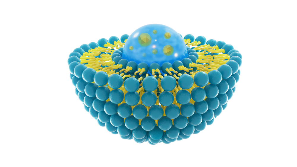
JNCL is caused by a mutation in the CLN3 gene, which provides instructions to make a lysosomal protein called battenin. This protein is found mostly in membranes surrounding lysosomes and endosomes — small compartments within cells that accumulate, digest, and recycle materials — but its precise function remains unknown.
Researchers at Sanford Children’s Health Research Center in Sioux Falls, South Dakota, may have finally identified one of its functions: to regulate the activity of cathepsin D (CTSD), a lysosomal enzyme whose malfunction leads to the accumulation of lipofuscins.
Previously, the same team had shown that mouse cells genetically engineered to produce an artificial form of the CLN3 protein significantly increased CLN3 production in response to certain conditions, specifically when the interior of these cells contained a higher concentration of dissolved materials compared to the exterior (osmolarity).
“Nevertheless, this past study did not clarify if the change in CLN3 subcellular distribution was a consequence of increased osmolarity or the increased CLN3 expression, and did not examine the possible lysosomal functions associated to the increased CLN3 expression,” the researchers wrote.
They’ve now found that both the artificial form of the CLN3 protein and cathepsin D cleaved to the membranes of lysosomes.
Importantly, they found the higher the production of CLN3 protein, the lower the activity of cathepsin D, suggesting that CLN3 could be directly controlling the activity of CTSD.
“Our results indicate a role of CLN3 in the regulation of CTSD activity. Under physiological, isotonic conditions, [mouse] cells with either transient or stable [CLN3] expression had significantly decreased CTSD enzyme activity,” researchers wrote.


Leave a comment
Fill in the required fields to post. Your email address will not be published.