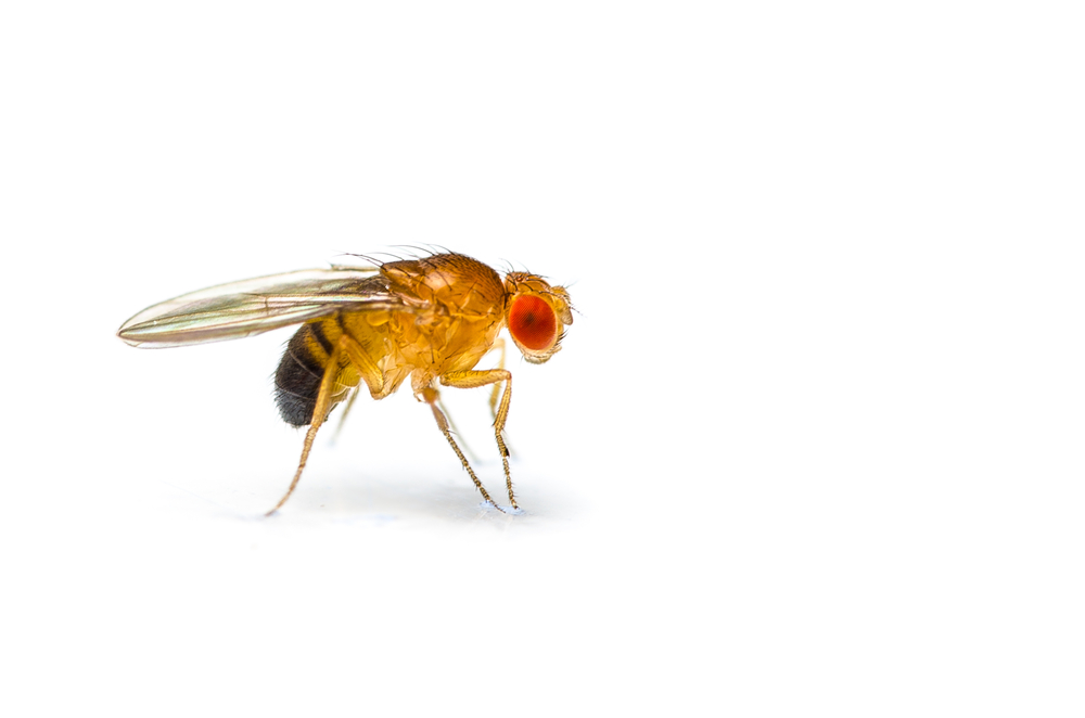
Fruit Fly Research Examines Two Genes Involved in Batten Disease

The expression of two genes associated with neuronal ceroid lipofuscinosis, better known as Batten disease, was restricted to certain cells of the central nervous system in a fruit fly research model.
Namely, these genes are mainly expressed in cells that form the blood brain barrier — a membrane that protects the brain from blood circulation — along with nerve cells involved in the development of vision.
The study, “in vivo localization of the neuronal ceroid lipofuscinosis proteins, CLN3 and CLN7, at endogenous expression levels,” was published in the journal Neurobiology of Disease.
The neuronal ceroid-lipofuscinoses (NCLs) are a group of inherited, neurodegenerative, lysosomal storage disorders characterized by progressive intellectual and motor deterioration, seizures, and early death. Vision loss is a feature of most forms. Researchers have identified 14 different genes responsible for NCL with varying ages of onset.
CLN3 disease, also called classic juvenile Batten disease, is one of the most prevalent forms of NCLs, with disease onset occurring between ages four and 10 years. Mutations in the CLN3 gene, which gives origin to a protein that is found not only in humans but also in simpler organisms like the fruit fly, account for this form of the disease.
Several lines of evidence point for a role of CLN3 in the epithelium (the tissue composed of cells that cover all body surfaces, line body cavities and hollow organs) in several organs. A mouse study also showed a strong pattern of CLN3 gene expression — the process by which information in a gene is synthesized to create a working product, like a protein — in the central nervous system (CNS). It is detected mainly in the later stages of embryonic development and persists after birth.
CLN7 is another form of Batten disease that typically begins between ages two and seven. While symptoms are similar to those of CLN3, the mutated gene behind this condition is called CLN7/MFS-domain containing 8 (CLN7/MFSD8) and gives rise to a protein localized in lysosomes, which are small vesicles involved in cell recycling that are affected in Batten disease.
The fruit fly Drosophila expresses only a subset of the CLN genes, which researchers hypothesized was likely to have core functions conserved in vertebrates.
The team used a gene editing tool called CRISPR/Cas9 to generate flies that have a yellow fluorescent protein, called YFP, fused to both CLN7 and CLN3, allowing them to visually observe where these two proteins are located within tissues.
CRISPR/Cas9 is a unique technology that enables researchers to edit parts of the genome by removing, adding or altering sections of the DNA sequence. Much like a tailor-made suit, genes can be “designed” to adequately and specifically serve researchers’ needs.
Mainly focusing on the CNS of Drosophila larvae, as Batten’s is largely restricted to this particular system, the team observed that both CLN3 and CLN7 proteins were largely restricted to subsets of glia cells forming the blood brain barrier in the CNS, with expression only in specific neurons.
Glia cells are the most abundant cells in the CNS and provide support to neurons by surrounding them and holding them in place. These cells also supply neurons with nutrients and oxygen, destroy microbes and remove dead neurons.
Although the CLN7 gene was mainly expressed within glia, certain neurons of the developing visual system also expressed this gene, “consistent with the early degeneration of the visual system in NCL patients,” researchers wrote.
CLN3 expression also was observed in glia cells and very strongly in Malpighian tubules — the fly equivalent of our kidneys. A closer look revealed the protein was present in microvilli (small protrusions on the cell’s surface), mitochondria (the cells’ powerhouses), and lysosomes within the Malpighian tubules.
“Surprisingly, we found that both CLN7 and CLN3 have restricted expression in the CNS: CLN7 is expressed in neurons in the developing visual system, but not widely in neurons elsewhere, and CLN3 is absent from neurons,” researchers wrote.
“Unexpectedly, we find that CLN3 is additionally localized at the apical domain of tubules and in mitochondria,” they added.
The authors believe that because CLN7 and CLN3 gene expression seems largely restricted to glia cells in the Drosophila CNS and the fact that activation of glial cells is the earliest detectable event in animal models of various NCLs, supports the use of Drosophila to gain insight into early developmental events underlying Batten’s disease.


