
Brain Cell Function Impaired in Early Forms of Batten Disease, Mouse Study Shows
Written by |


Glial cell function is impaired in the juvenile form of Batten disease, leading to nerve cell loss, a mouse study has found.
The study, “Glial cells are functionally impaired in juvenile neuronal ceroid lipofuscinosis and detrimental to neurons,” was appeared in the journal Acta Neuropathologica Communications.
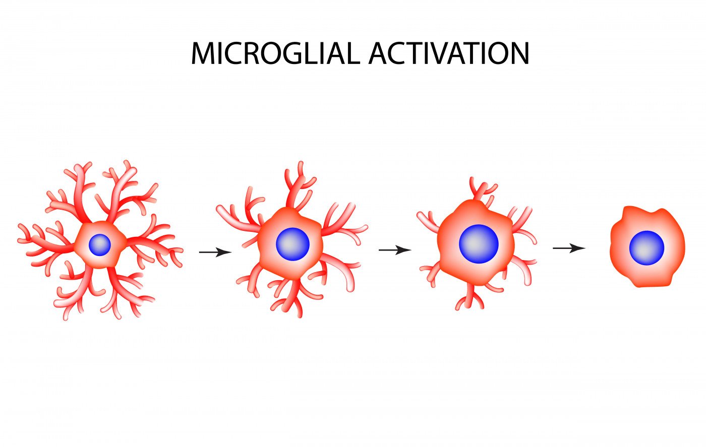
Glial cells, a term that comprises many types of cells, such as microglia, astrocytesa and oligodendrocytes, make up much of the mammalian brain. Doctors have observed localized activation of glial cells in Batten disease from early on. Moreover, the regions where glial cells are activated seem to accurately predict where neuron loss will occur. This is particularly true for astrocytes and microglia activation, as human autopsy studies have suggested.
In the most common juvenile form of Batten, called JNCL or CLN3 disease, several studies have suggested that the activation of both astrocytes and microglia is higher when compared to other earlier onset forms of Batten.
Researchers investigating this phenomenon further have confirmed that in both mouse models and human autopsy material, the glial response is less pronounced. They isolated glial cells and neurons from a mouse model of juvenile Batten disease (the Cln3-deficient mice) and tested their properties in vitro.
The isolated cells, after stimulation, failed to transform their shape; their secretion abilities were also attenuated. This was particularly evident for astrocytes that exhibited impaired secretion of several neuroprotective factors, and other important signaling molecules.
“Most importantly, using a co-culture system, Cln3-deficient astrocytes and microglia had a negative impact on the survival and morphology of both Cln3-deficient and wild-type neurons,” authors wrote.
These results suggest that in juvenile Batten disease the function of glial cells – particularly, astrocytes – is compromised. Together with impaired microglia cells, their loss of function seems to promote loss of nerve cells.
Overall, these results “provide further new information on how both glia and neurons are compromised in this disorder and the negative role that glial cells appear to play in the pathogenesis of CLN3 disease,” researchers wrote.

Leave a comment
Fill in the required fields to post. Your email address will not be published.