
Researchers Dissect Mechanisms Involved in CLN5 Release Using Amoeba Model


Using a species of amoeba as a model organism, researchers have found chemical modifications in the CLN5 protein — whose mutated form is associated with late infantile Batten disease — that allow it to be released outside a cell.
According to the team from Trent University in Canada, their findings provide evidence that a similar mechanism in humans could be involved in disease onset and progression.
The study, “Secretion and function of Cln5 during the early stages of Dictyostelium development,” was published in Biochimica et Biophysica Acta (BBA) – Molecular Research.
Neuronal ceroid lipofuscinoses (NCLs), also known as Batten disease, comprise a group of childhood genetic neurodegenerative disorders with a range of symptoms, including vision loss, lack of motor coordination, and impaired cognition.
These conditions can be caused by mutations in 14 different genes (CLN1 to CLN14), that lead to the accumulation of toxic, insoluble waste deposits, called lipofuscins, inside cells. One such gene is CLN5, which has been associated with the late infantile form of the disease, also known as neuronal ceroid lipofuscinosis 5 (CLN5-NCL).
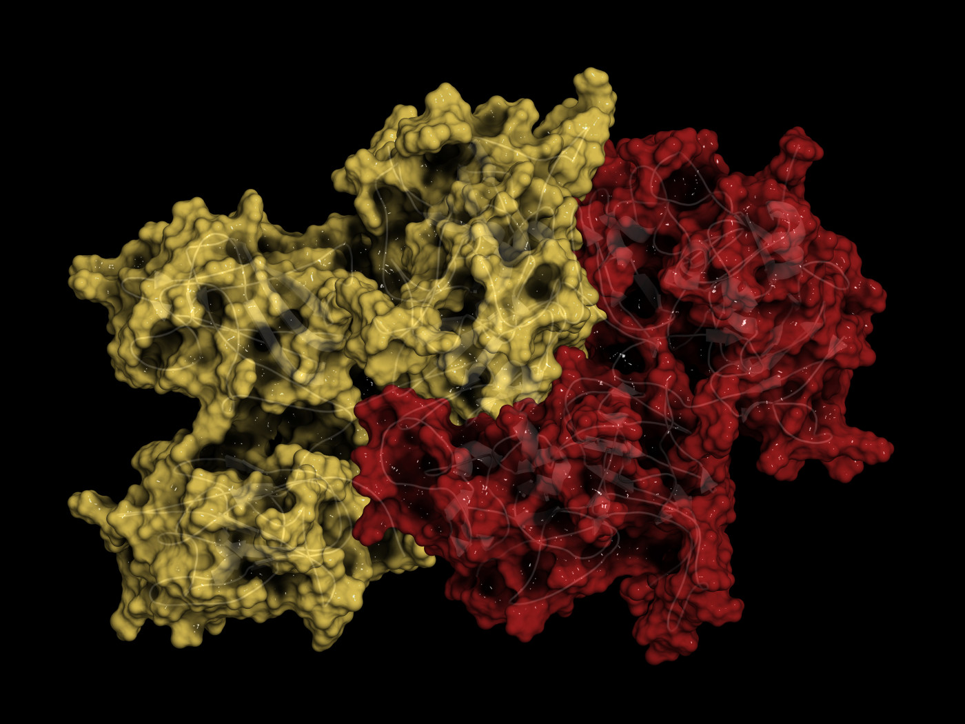
This gene provides instructions for the production of the CLN5 protein, whose function is still poorly understood. Once active, CLN5 is transported to lysosomes — special compartments within cells that digest and recycle different types of molecules — where it is thought to regulate the transit of recycled materials between different cell compartments.
Several genetic models of disease have been created in an attempt to study the function of CLN5, but this field of research has been hindered by the lack of simple eukaryotic (organisms whose cells have a nucleus surrounded by a membrane) model systems that contain a protein equivalent to the human CLN5.
In a previous study, the same team found a species of amoeba, called Dictyostelium discoideum, that produced a protein similar to human CLN5. Importantly, their findings also revealed this protein worked as a glycoside hydrolase — an enzyme that breaks down complex sugar molecules — and localized to the endoplasmatic reticulum, a compartment within cells responsible for molecule trafficking, before being released.
In this study, the group created an antibody that specifically recognizes Dictyostelium‘s CLN5 to study in more detail how this protein is released to the outside of cells during the early stages of amoeba development.
Like human CLN5, the protein found in amoebas requires a special chemical modification, called glycosylation, to be released to the outside. However, this protein seems to follow an unconventional pathway for its release, completely bypassing the Golgi complex (another cell compartment involved in molecule trafficking) and instead using a pathway linked to autophagy, the process of controlled cell degradation and recycling.
Interestingly, researchers also found that CLN5 co-localized with CLN3 — a protein associated with the juvenile form of Batten disease in humans, also known as CLN3 disease — in Dictyostelium and noticed a higher release of CLN5 in genetically modified cells unable to produce CLN3, suggesting that CLN3 regulates CLN5 release in this model organism.
In addition, investigators found that silencing CLN5 led to the same kind of cell adhesion and chemotaxis (the ability of cells to detect and move toward a chemical substance) defects present in Dictyostelium cells lacking CLN3.
“Together, our data support a function for CLN5 during the early stages of multicellular development, provide further evidence for the molecular networking of NCL proteins, and provide insight into the mechanisms that may underlie CLN5 function in humans,” the researchers wrote.